ServiceAvailable
H&E and Special Histological Stains
Lady Davis Institute
Core facility
Jewish General Hospital
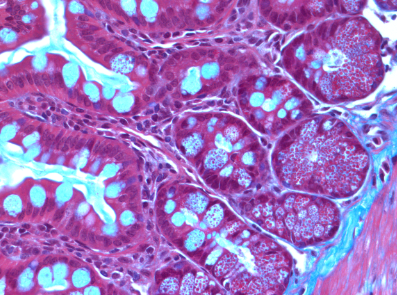
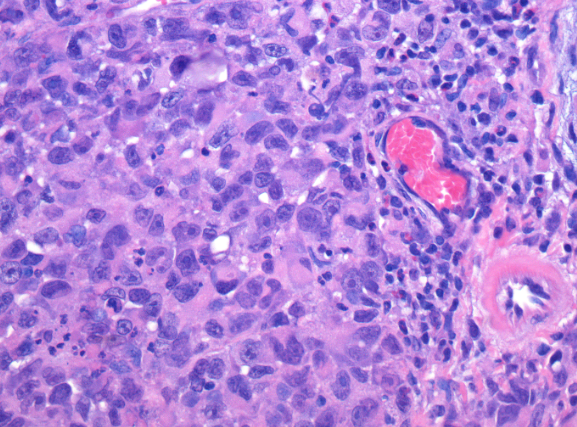
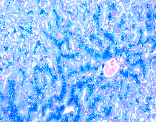
This service provides a wide range of histological staining techniques to visualize cellular and tissue structures. Hematoxylin and Eosin (H&E) staining is the most common and fundamental method, using hematoxylin to stain cell nuclei blue/purple and eosin to stain cytoplasm and extracellular matrix components pink/red, providing a comprehensive overview of tissue microanatomy and morphology. Additionally, a variety of special stains are offered to highlight specific tissue components or pathologies. Examples include: Masson Trichrome (for collagen and muscle fibers), Perl’s Iron (for iron deposits), PAS (Periodic Acid-Schiff, for carbohydrates like glycogen and mucins), Sirius Red (for collagen, especially in fibrosis), Luxol Fast Blue (for myelin), Von Kossa (for calcium deposits), Oil-O-Red (for lipids), and Giemsa (for blood cells, bacteria, and parasites). These specialized stains are crucial for detailed diagnostic evaluation and research applications, revealing subtle nuances not visible with H&E alone.
The George and Olga Minarik Research Pathology Facility
Lady Davis Institute
Research lab focused on advancing scientific knowledge and innovation.
NB
Naciba Benlimame
ServiceAvailable
H&E and Special Histological Stains
Lady Davis Institute
Core facility
Jewish General Hospital
This service provides a wide range of histological staining techniques to visualize cellular and tissue structures. Hematoxylin and Eosin (H&E) staining is the most common and fundamental method, using hematoxylin to stain cell nuclei blue/purple and eosin to stain cytoplasm and extracellular matrix components pink/red, providing a comprehensive overview of tissue microanatomy and morphology. Additionally, a variety of special stains are offered to highlight specific tissue components or pathologies. Examples include: Masson Trichrome (for collagen and muscle fibers), Perl’s Iron (for iron deposits), PAS (Periodic Acid-Schiff, for carbohydrates like glycogen and mucins), Sirius Red (for collagen, especially in fibrosis), Luxol Fast Blue (for myelin), Von Kossa (for calcium deposits), Oil-O-Red (for lipids), and Giemsa (for blood cells, bacteria, and parasites). These specialized stains are crucial for detailed diagnostic evaluation and research applications, revealing subtle nuances not visible with H&E alone.
The George and Olga Minarik Research Pathology Facility
Lady Davis Institute
Research lab focused on advancing scientific knowledge and innovation.
NB
Naciba Benlimame
You might also like
Discover more resources that could support your research