EquipmentAvailable
CIAN-SP8 Confocal Laser Scanning Microscope
Faculty of Science
Core Facility
McGill University
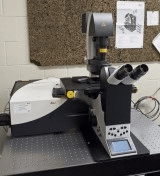
The Leica SP8 is a point-scanning confocal system mounted on a fully motorized Leica DMI6000B inverted microscope. It is equipped with a "SuperZ" Galvo stage for precise focusing and z-stack acquisition. The system includes four spectral fluorescence detectors—three photomultiplier tubes (PMTs) and one high-sensitivity Hybrid Detector (HyD)—along with a transmitted light detector (PMT).
Objectives:
- 10x/0.4 Dry: HC PL APO 10x/0.40 CS
- 20x/0.7 Dry: HC PL APO 20x/0.70 CS
- 40x/0.85 Dry: HCX PL APO 40x/0.85 CORR CS
- 40x/1.1 Water: HC PL APO 40x/1.10 W CORR CS2
- 40x/1.3 Oil: HC PL APO 40x/1.30 Oil CS2
- 63x/1.4 Oil: HC PL APO 63x/1.40 Oil CS2 McGill University
Laser Lines:
- 405 nm Diode (50 mW): Excitation for DAPI
- 448 nm Diode (40 mW): Excitation for CFP
- 488 nm Diode (20 mW): Excitation for AF488, GFP, YFP
- 552 nm Diode (20 mW): Excitation for TRITC, Cy3, Texas Red, RFP
- 638 nm Diode (30 mW): Excitation for AF633, Cy5, FarRedFP McGill University
Features:
- Spectral Detection: Tunable emission detection without the need for emission filters, allowing flexibility in fluorophore selection.
- Wide-Field Fluorescence: Equipped with a Leica EL6000 metal-halide lamp and filter sets for DAPI, GFP, and RFP.
- Differential Interference Contrast (DIC): Available for high-contrast imaging of transparent specimens.
This system is ideal for high-resolution imaging applications, including multi-color fluorescence, live-cell imaging, and spectral unmixing. It is located within the Cell Imaging and Analysis Network (CIAN) at McGill University.
Initial training sessions usually last about 3 hours and include the hourly microscope fee plus the technical support fee.
IMPORTANT: ALL training and technical support is by appointment ONLY and will depend on the availability of the ABIF staff.
Advanced BioImaging Facility
Faculty of Science
Research lab focused on advancing scientific knowledge and innovation.
CB
Claire Brown
EquipmentAvailable
CIAN-SP8 Confocal Laser Scanning Microscope
Faculty of Science
Core Facility
McGill University
The Leica SP8 is a point-scanning confocal system mounted on a fully motorized Leica DMI6000B inverted microscope. It is equipped with a "SuperZ" Galvo stage for precise focusing and z-stack acquisition. The system includes four spectral fluorescence detectors—three photomultiplier tubes (PMTs) and one high-sensitivity Hybrid Detector (HyD)—along with a transmitted light detector (PMT).
Objectives:
- 10x/0.4 Dry: HC PL APO 10x/0.40 CS
- 20x/0.7 Dry: HC PL APO 20x/0.70 CS
- 40x/0.85 Dry: HCX PL APO 40x/0.85 CORR CS
- 40x/1.1 Water: HC PL APO 40x/1.10 W CORR CS2
- 40x/1.3 Oil: HC PL APO 40x/1.30 Oil CS2
- 63x/1.4 Oil: HC PL APO 63x/1.40 Oil CS2 McGill University
Laser Lines:
- 405 nm Diode (50 mW): Excitation for DAPI
- 448 nm Diode (40 mW): Excitation for CFP
- 488 nm Diode (20 mW): Excitation for AF488, GFP, YFP
- 552 nm Diode (20 mW): Excitation for TRITC, Cy3, Texas Red, RFP
- 638 nm Diode (30 mW): Excitation for AF633, Cy5, FarRedFP McGill University
Features:
- Spectral Detection: Tunable emission detection without the need for emission filters, allowing flexibility in fluorophore selection.
- Wide-Field Fluorescence: Equipped with a Leica EL6000 metal-halide lamp and filter sets for DAPI, GFP, and RFP.
- Differential Interference Contrast (DIC): Available for high-contrast imaging of transparent specimens.
This system is ideal for high-resolution imaging applications, including multi-color fluorescence, live-cell imaging, and spectral unmixing. It is located within the Cell Imaging and Analysis Network (CIAN) at McGill University.
Initial training sessions usually last about 3 hours and include the hourly microscope fee plus the technical support fee.
IMPORTANT: ALL training and technical support is by appointment ONLY and will depend on the availability of the ABIF staff.
Advanced BioImaging Facility
Faculty of Science
Research lab focused on advancing scientific knowledge and innovation.
CB
Claire Brown
You might also like
Discover more resources that could support your research