EquipmentAvailable
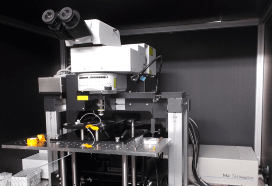
Olympus FV1000MPE multiphoton and laser scanning confocal microscope
Faculty of Medicine and Health Sciences
Research Institute of the McGill University Health Centre
McGill University
Specifications
Microscopy stand: Upright BX61WI microscope with motorized XY stage and portal frame for in vivo imaging
Software: Fluoview Advanced Software
Detector:
- Four (4) channel non-descanned MPE detection
- Three (3) channel visible confocal detection with two (2) of the detectors being spectral
- One (1) transmitted light detector for visible laser DIC
Lasers:
- 405 nm, 440 nm, 488 nm, 514 nm, 561 nm and 635 nm
- Mai Tai HP DeepSee laser (tunable IR laser from 690-1040 nm)
- Dual output enables use of all laser lines for imaging and photo-manipulation
- Live cell module: CO2 and temperature controller
Objective:
- Plan-Neofluar 20x LD
Applications
Multi-fluorescence with DIC
3D examinations (Z-stacks)
Multidimensional acquisition (tile scan, time lapse)
Confocal imaging
Intravital imaging
Live cell imaging
FRET
Photo-manipulation (FRAP, uncaging, etc.)
Molecular Imaging Platform - RI-MUHC
Faculty of Medicine and Health Sciences
Research lab focused on advancing scientific knowledge and innovation.
MF
Min Fu
EquipmentAvailable
Olympus FV1000MPE multiphoton and laser scanning confocal microscope
Faculty of Medicine and Health Sciences
Research Institute of the McGill University Health Centre
McGill University
Specifications
Microscopy stand: Upright BX61WI microscope with motorized XY stage and portal frame for in vivo imaging
Software: Fluoview Advanced Software
Detector:
- Four (4) channel non-descanned MPE detection
- Three (3) channel visible confocal detection with two (2) of the detectors being spectral
- One (1) transmitted light detector for visible laser DIC
Lasers:
- 405 nm, 440 nm, 488 nm, 514 nm, 561 nm and 635 nm
- Mai Tai HP DeepSee laser (tunable IR laser from 690-1040 nm)
- Dual output enables use of all laser lines for imaging and photo-manipulation
- Live cell module: CO2 and temperature controller
Objective:
- Plan-Neofluar 20x LD
Applications
Multi-fluorescence with DIC
3D examinations (Z-stacks)
Multidimensional acquisition (tile scan, time lapse)
Confocal imaging
Intravital imaging
Live cell imaging
FRET
Photo-manipulation (FRAP, uncaging, etc.)
Molecular Imaging Platform - RI-MUHC
Faculty of Medicine and Health Sciences
Research lab focused on advancing scientific knowledge and innovation.
MF
Min Fu
You might also like
Discover more resources that could support your research