EquipmentAvailable
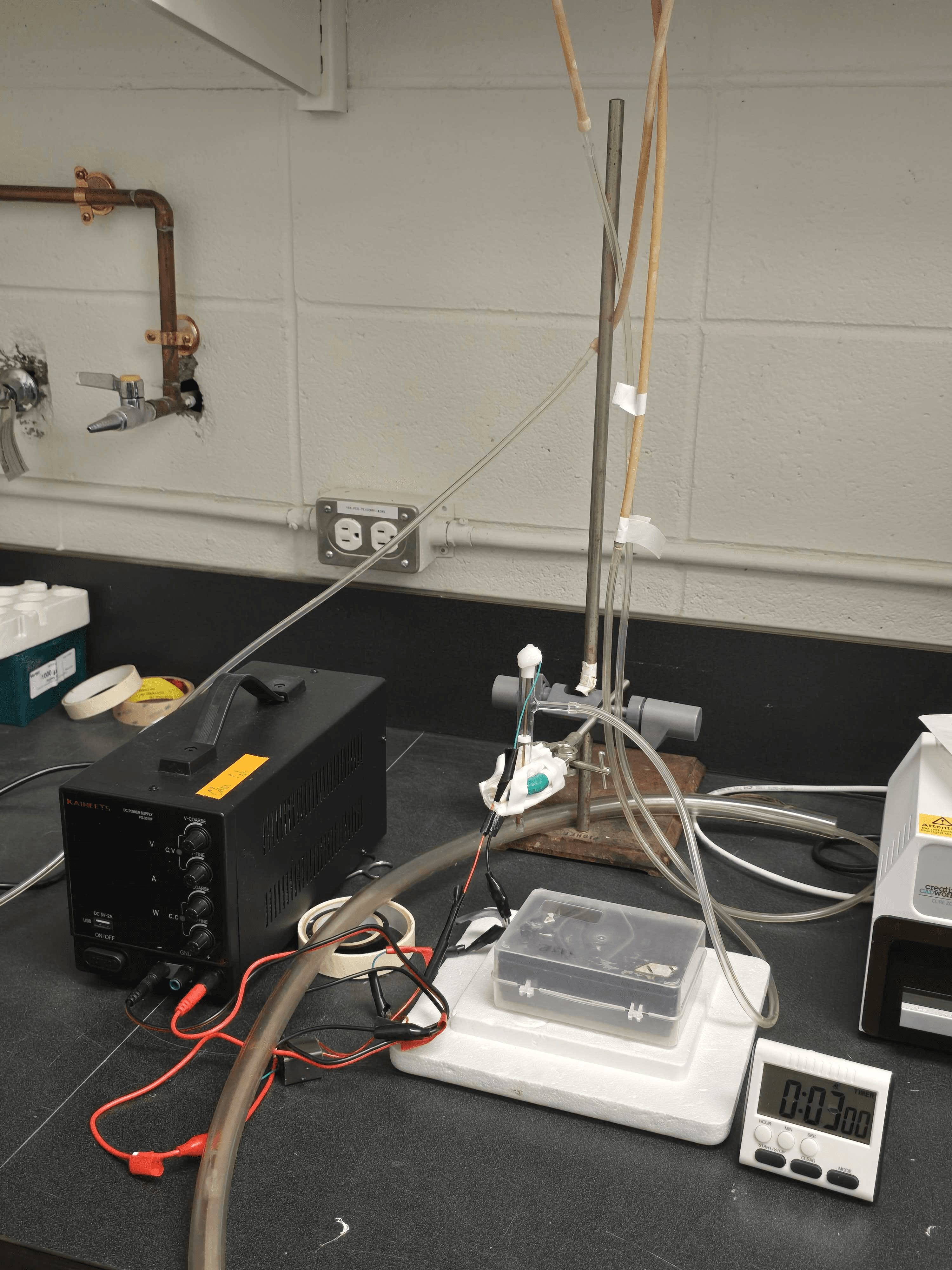
Custom Cold Atmospheric Plasma–Hydrogel Activation Station
Faculty of Medicine and Health Sciences
Biomedical Engineering
McGill University
Proprietary platform for generating and preserving reactive plasma species in injectable hydrogels
Purpose & Applications
This benchtop setup is specifically designed to activate hydrogels with cold atmospheric plasma (CAP), allowing the hydrogel network to “store” therapeutically active reactive oxygen and nitrogen species (RONS). In Dr. Guojun Chen’s work (e.g., Biomaterials 2023, Proc. Natl. Acad. Sci. 2020), these plasma‐activated immunotherapeutic hydrogels are subsequently injected or applied to tumor beds to:
- Enhance cancer immunotherapy by delivering a sustained bolus of ROS/RNS in situ
- Synergize with immune checkpoint blockade (ICB), improving local and systemic anti‐tumor immune responses
- Achieve localized tumor killing and minimal off‐target toxicity, since the reactive species are preserved within the gel matrix until injection
Beyond cancer applications, this platform can also be adapted for:
- Sterilization or surface crosslinking of protein‐ or polysaccharide‐based gels
- Tuning mechanical properties of hydrogel scaffolds via controlled oxidative crosslinking
- Embedding RONS for wound‐healing or antimicrobial gels
Core Components & Workflow (High‐Level)
- Cold Plasma Generator
- Helium/Nitrogen Feed:
- • High‐purity helium (≥ 99.99 %) or nitrogen is delivered (typical flow 300–500 sccm) through a narrow‐bore stainless‐steel nozzle.
- • Low‐flow O₂ admixture (1–2 %) can be introduced to boost RONS production if needed.
- DC Power Supply:
- • Provides a stable 10–20 V DC across a fine tungsten needle (anode) and a grounded counter‐electrode.
- • Current is adjusted (~ 0.2 A–0.8 A) to maintain a uniform, non‐thermal glow discharge just above the gel surface.
- Electrode Assembly:
- • A pointed tungsten or stainless‐steel needle is positioned ~ 1–3 mm above the gel precursor.
- • The ground electrode (stainless‐steel plate or mesh) sits beneath or adjacent to the gel container, completing the circuit.
- • Insulating ceramic spacers and PTFE collars ensure that the plasma discharge remains localized to the gel interface.
- Hydrogel Preparation & Positioning
- Gel Precursors:
- • Common formulations include Pluronic F127, collagen/fibrin blends, or thermosensitive polymer solutions that will gel upon injection.
- • Precursors are cast into shallow dishes (20–30 mm diameter, 5–10 mm depth) on a Teflon or PTFE sample stage.
- Stage & Height Adjustment:
- • The gel dish sits on a foam‐insulated block to prevent heat accumulation during plasma exposure.
- • A manual vertical adjuster (retort rod with plastic clips) aligns the electrode tip ~ 2 mm above the gel surface.
- Plasma Activation Protocol
- Power & Gas Initialization:
- • Power supply is switched to constant‐voltage (CV) mode at ~ 15 V; current limit set to ~ 0.5 A.
- • Helium (or He/N₂) flow is ramped to ~ 300 sccm; stable plasma plume (~ 3 mm length) ignites at the needle tip.
- Treatment Duration:
- • Typical exposure times range from 30 s to 2 min, depending on desired RONS load.
- • For immunotherapeutic hydrogels (per Chen et al.), a 60 s continuous treatment was found optimal to load long‐lived ROS/RNS without overheating the gel.
- Uniformity:
- • The electrode is scanned gently over the gel surface (± 5 mm) or held stationary at the center—either approach delivers a homogenous RONS distribution.
- • A small petri‐dish‐sized ground electrode (beneath the stage) ensures even coverage.
- Post‐Activation Handling
- Immediate Transfer:
- • Once plasma treatment concludes, the gel is capped or covered (petri dish lid) within 30 s to minimize ambient neutralization of RONS.
- • Activated gel can be kept at 4 °C (ice bath) for up to 30 min with minimal loss of reactive species.
- Injection or Application:
- • For in vivo studies (e.g., intratumoral injections), the RONS‐laden gel is drawn into syringes (27–30 G needle) and administered immediately (within 10 min of activation).
- • For in vitro assays (e.g., cell‐seeding on plasma‐treated collagen gels), the gel is transferred to culture plates under sterile conditions.
Key Applications in Dr. Chen’s Publications
- Injectable Plasma‐Activated Immunotherapeutic Hydrogel (Biomaterials 2023)
- • Pluronic F127 hydrogel treated with CAP retains long‐lived ROS/RNS (e.g., hydrogen peroxide, nitrites) that remain active for ≥ 30 min.
- • When co‐delivered with anti‐PD‐1 antibodies, the CAP‐activated gel amplifies local tumor antigen release and primes systemic immunity.
- Transdermal Plasma‐Mediated Immune Checkpoint Blockade (PNAS 2020)
- • Although primarily a skin application, the same CAP generator concept was used to “pre‐charge” a thermoresponsive gel that adhered to post‐surgical tumor beds, releasing RONS over several hours.
- Portable Plasma Jet for Postsurgical Ablation (Sci. Adv. 2021)
- • The benchtop plasma source was adapted into a handheld design; the gel activation station remains the “gold standard” for preparing RONS‐loaded gels in a sterile, controlled bench environment.
- Plasma‐Activated Biogel for Postoperative Therapy (Biomaterials 2021)
- • A hyaluronic acid–based gel was plasma‐treated to produce surface‐bound radicals, then directly applied to wound sites to promote wound healing and sterilization.
Why This Setup Matters
- Preserves Reactive Species:
- By tailoring the distance, gas flow, and exposure time, the system “locks” RONS into the gel network, extending their half‐life compared to direct plasma exposure.
- Versatile Hydrogel Compatibility:
- Compatible with a wide range of gel formulations—thermosensitive, photocurable, or enzymatically crosslinking systems.
- Rapid Prototyping & Iteration:
- Researchers can test multiple gel compositions, plasma gas mixtures (He vs. N₂ vs. O₂ admixture), and exposure times in a single afternoon to quickly optimize RONS delivery.
- Sterile & Enclosed:
- The entire rig sits on a standard biosafety bench, and all surfaces can be wiped with ethanol—enabling aseptic plasma‐gel preparation for cell culture or in vivo studies.
- Broad Biomedical Impact:
- Beyond oncology, similar CAP‐hydrogel constructs have been used for antimicrobial wound dressings, antifibrotic gel coatings, and surface functionalization of 3D cell‐culture scaffolds.
In summary, this custom CAP–hydrogel activation station is central to Dr. Guojun Chen’s work on plasma‐enhanced immunotherapeutic hydrogels. By generating a stable, low‐temperature glow discharge directly over liquid or semi‐solid gel precursors, it embeds long‐lived reactive species into the material—enabling injectable or surface‐bound RONS delivery for cancer therapy, wound healing, and advanced biomaterials research.
Chen Lab
Faculty of Medicine and Health Sciences
Research lab focused on advancing scientific knowledge and innovation.
GC
Guojun Chen
EquipmentAvailable
Custom Cold Atmospheric Plasma–Hydrogel Activation Station
Faculty of Medicine and Health Sciences
Biomedical Engineering
McGill University
Proprietary platform for generating and preserving reactive plasma species in injectable hydrogels
Purpose & Applications
This benchtop setup is specifically designed to activate hydrogels with cold atmospheric plasma (CAP), allowing the hydrogel network to “store” therapeutically active reactive oxygen and nitrogen species (RONS). In Dr. Guojun Chen’s work (e.g., Biomaterials 2023, Proc. Natl. Acad. Sci. 2020), these plasma‐activated immunotherapeutic hydrogels are subsequently injected or applied to tumor beds to:
- Enhance cancer immunotherapy by delivering a sustained bolus of ROS/RNS in situ
- Synergize with immune checkpoint blockade (ICB), improving local and systemic anti‐tumor immune responses
- Achieve localized tumor killing and minimal off‐target toxicity, since the reactive species are preserved within the gel matrix until injection
Beyond cancer applications, this platform can also be adapted for:
- Sterilization or surface crosslinking of protein‐ or polysaccharide‐based gels
- Tuning mechanical properties of hydrogel scaffolds via controlled oxidative crosslinking
- Embedding RONS for wound‐healing or antimicrobial gels
Core Components & Workflow (High‐Level)
- Cold Plasma Generator
- Helium/Nitrogen Feed:
- • High‐purity helium (≥ 99.99 %) or nitrogen is delivered (typical flow 300–500 sccm) through a narrow‐bore stainless‐steel nozzle.
- • Low‐flow O₂ admixture (1–2 %) can be introduced to boost RONS production if needed.
- DC Power Supply:
- • Provides a stable 10–20 V DC across a fine tungsten needle (anode) and a grounded counter‐electrode.
- • Current is adjusted (~ 0.2 A–0.8 A) to maintain a uniform, non‐thermal glow discharge just above the gel surface.
- Electrode Assembly:
- • A pointed tungsten or stainless‐steel needle is positioned ~ 1–3 mm above the gel precursor.
- • The ground electrode (stainless‐steel plate or mesh) sits beneath or adjacent to the gel container, completing the circuit.
- • Insulating ceramic spacers and PTFE collars ensure that the plasma discharge remains localized to the gel interface.
- Hydrogel Preparation & Positioning
- Gel Precursors:
- • Common formulations include Pluronic F127, collagen/fibrin blends, or thermosensitive polymer solutions that will gel upon injection.
- • Precursors are cast into shallow dishes (20–30 mm diameter, 5–10 mm depth) on a Teflon or PTFE sample stage.
- Stage & Height Adjustment:
- • The gel dish sits on a foam‐insulated block to prevent heat accumulation during plasma exposure.
- • A manual vertical adjuster (retort rod with plastic clips) aligns the electrode tip ~ 2 mm above the gel surface.
- Plasma Activation Protocol
- Power & Gas Initialization:
- • Power supply is switched to constant‐voltage (CV) mode at ~ 15 V; current limit set to ~ 0.5 A.
- • Helium (or He/N₂) flow is ramped to ~ 300 sccm; stable plasma plume (~ 3 mm length) ignites at the needle tip.
- Treatment Duration:
- • Typical exposure times range from 30 s to 2 min, depending on desired RONS load.
- • For immunotherapeutic hydrogels (per Chen et al.), a 60 s continuous treatment was found optimal to load long‐lived ROS/RNS without overheating the gel.
- Uniformity:
- • The electrode is scanned gently over the gel surface (± 5 mm) or held stationary at the center—either approach delivers a homogenous RONS distribution.
- • A small petri‐dish‐sized ground electrode (beneath the stage) ensures even coverage.
- Post‐Activation Handling
- Immediate Transfer:
- • Once plasma treatment concludes, the gel is capped or covered (petri dish lid) within 30 s to minimize ambient neutralization of RONS.
- • Activated gel can be kept at 4 °C (ice bath) for up to 30 min with minimal loss of reactive species.
- Injection or Application:
- • For in vivo studies (e.g., intratumoral injections), the RONS‐laden gel is drawn into syringes (27–30 G needle) and administered immediately (within 10 min of activation).
- • For in vitro assays (e.g., cell‐seeding on plasma‐treated collagen gels), the gel is transferred to culture plates under sterile conditions.
Key Applications in Dr. Chen’s Publications
- Injectable Plasma‐Activated Immunotherapeutic Hydrogel (Biomaterials 2023)
- • Pluronic F127 hydrogel treated with CAP retains long‐lived ROS/RNS (e.g., hydrogen peroxide, nitrites) that remain active for ≥ 30 min.
- • When co‐delivered with anti‐PD‐1 antibodies, the CAP‐activated gel amplifies local tumor antigen release and primes systemic immunity.
- Transdermal Plasma‐Mediated Immune Checkpoint Blockade (PNAS 2020)
- • Although primarily a skin application, the same CAP generator concept was used to “pre‐charge” a thermoresponsive gel that adhered to post‐surgical tumor beds, releasing RONS over several hours.
- Portable Plasma Jet for Postsurgical Ablation (Sci. Adv. 2021)
- • The benchtop plasma source was adapted into a handheld design; the gel activation station remains the “gold standard” for preparing RONS‐loaded gels in a sterile, controlled bench environment.
- Plasma‐Activated Biogel for Postoperative Therapy (Biomaterials 2021)
- • A hyaluronic acid–based gel was plasma‐treated to produce surface‐bound radicals, then directly applied to wound sites to promote wound healing and sterilization.
Why This Setup Matters
- Preserves Reactive Species:
- By tailoring the distance, gas flow, and exposure time, the system “locks” RONS into the gel network, extending their half‐life compared to direct plasma exposure.
- Versatile Hydrogel Compatibility:
- Compatible with a wide range of gel formulations—thermosensitive, photocurable, or enzymatically crosslinking systems.
- Rapid Prototyping & Iteration:
- Researchers can test multiple gel compositions, plasma gas mixtures (He vs. N₂ vs. O₂ admixture), and exposure times in a single afternoon to quickly optimize RONS delivery.
- Sterile & Enclosed:
- The entire rig sits on a standard biosafety bench, and all surfaces can be wiped with ethanol—enabling aseptic plasma‐gel preparation for cell culture or in vivo studies.
- Broad Biomedical Impact:
- Beyond oncology, similar CAP‐hydrogel constructs have been used for antimicrobial wound dressings, antifibrotic gel coatings, and surface functionalization of 3D cell‐culture scaffolds.
In summary, this custom CAP–hydrogel activation station is central to Dr. Guojun Chen’s work on plasma‐enhanced immunotherapeutic hydrogels. By generating a stable, low‐temperature glow discharge directly over liquid or semi‐solid gel precursors, it embeds long‐lived reactive species into the material—enabling injectable or surface‐bound RONS delivery for cancer therapy, wound healing, and advanced biomaterials research.
Chen Lab
Faculty of Medicine and Health Sciences
Research lab focused on advancing scientific knowledge and innovation.
GC
Guojun Chen
You might also like
Discover more resources that could support your research