EquipmentAvailable
Human Research PET Imaging – ECAT High Resolution Research Tomograph (HRRT)
Faculty of Medicine and Health Sciences
Core Facility
McGill University
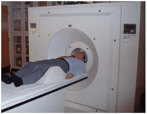
The ECAT High Resolution Research Tomograph (HRRT) is a dedicated human brain PET camera designed for high sensitivity and unmatched spatial resolution in neuroimaging. Installed at the BIC in 2007, the HRRT remains one of the most advanced PET scanners available, with only seventeen units worldwide, contributing to significant advancements in neuroscience research.
Key Features & Capabilities
🔹 Unmatched Spatial Resolution
- 2.4 mm resolution at the center of the Field of View (FoV), surpassing commercial human PET scanners.
- 119,808 small crystals (2.1 × 2.1 × 10 mm³) distributed across 8 detector panels in an octagonal design.
- Quadrant-sharing photomultiplier tubes (PMTs) enhance precision.
🔹 Optimized Field of View (FoV) for Whole-Head Imaging
- Bore opening: 35 cm diameter
- Axial FoV: 25 cm
- Radial FoV: 31.2 cm
🔹 Dual-Layer Crystal Configuration for Sensitivity & Depth Resolution
- Front layer: Lutetium Orthosilicate (LSO)
- Back layer: Lutetium Yttrium Orthosilicate (LYSO)
- Sensitivity of 4.3% at the FoV center, improving signal detection across brain structures.
🔹 Advanced Data Acquisition & Image Reconstruction
- List mode acquisition for post-processing flexibility in temporal sampling.
- Fast attenuation map acquisition using a Cs-137 source for transmission imaging.
- State-of-the-art image reconstruction with corrections for motion artifacts, partial volume effects, and PET degradation factors.
- In-house motion correction techniques to reduce image degradation caused by patient movement.
Applications
✅ Neuroscience Research – Mapping neurotransmitter systems, receptor binding, and metabolic activity in various brain disorders.
✅ Neurodegenerative Disease Studies – Investigating Alzheimer’s, Parkinson’s, and other neurodegenerative conditions.
✅ Pharmacokinetic Studies – Measuring drug distribution and receptor interactions in the brain.
✅ Cognitive and Behavioral Studies – Exploring brain function during cognitive tasks.
Access & Inquiries
For more details on Human Research PET Imaging, tracer availability, or research collaborations, please visit the BIC PET Imaging Unit or contact our expert PET analysis team.
The HRRT scanner offers unparalleled precision in human brain PET imaging, ensuring high-quality, quantitative, and reproducible neuroimaging data for cutting-edge research.
McConnell Brain Imaging Centre (BIC)
Faculty of Medicine and Health Sciences
Research lab focused on advancing scientific knowledge and innovation.
JD
Julien Doyon
EquipmentAvailable
Human Research PET Imaging – ECAT High Resolution Research Tomograph (HRRT)
Faculty of Medicine and Health Sciences
Core Facility
McGill University
The ECAT High Resolution Research Tomograph (HRRT) is a dedicated human brain PET camera designed for high sensitivity and unmatched spatial resolution in neuroimaging. Installed at the BIC in 2007, the HRRT remains one of the most advanced PET scanners available, with only seventeen units worldwide, contributing to significant advancements in neuroscience research.
Key Features & Capabilities
🔹 Unmatched Spatial Resolution
- 2.4 mm resolution at the center of the Field of View (FoV), surpassing commercial human PET scanners.
- 119,808 small crystals (2.1 × 2.1 × 10 mm³) distributed across 8 detector panels in an octagonal design.
- Quadrant-sharing photomultiplier tubes (PMTs) enhance precision.
🔹 Optimized Field of View (FoV) for Whole-Head Imaging
- Bore opening: 35 cm diameter
- Axial FoV: 25 cm
- Radial FoV: 31.2 cm
🔹 Dual-Layer Crystal Configuration for Sensitivity & Depth Resolution
- Front layer: Lutetium Orthosilicate (LSO)
- Back layer: Lutetium Yttrium Orthosilicate (LYSO)
- Sensitivity of 4.3% at the FoV center, improving signal detection across brain structures.
🔹 Advanced Data Acquisition & Image Reconstruction
- List mode acquisition for post-processing flexibility in temporal sampling.
- Fast attenuation map acquisition using a Cs-137 source for transmission imaging.
- State-of-the-art image reconstruction with corrections for motion artifacts, partial volume effects, and PET degradation factors.
- In-house motion correction techniques to reduce image degradation caused by patient movement.
Applications
✅ Neuroscience Research – Mapping neurotransmitter systems, receptor binding, and metabolic activity in various brain disorders.
✅ Neurodegenerative Disease Studies – Investigating Alzheimer’s, Parkinson’s, and other neurodegenerative conditions.
✅ Pharmacokinetic Studies – Measuring drug distribution and receptor interactions in the brain.
✅ Cognitive and Behavioral Studies – Exploring brain function during cognitive tasks.
Access & Inquiries
For more details on Human Research PET Imaging, tracer availability, or research collaborations, please visit the BIC PET Imaging Unit or contact our expert PET analysis team.
The HRRT scanner offers unparalleled precision in human brain PET imaging, ensuring high-quality, quantitative, and reproducible neuroimaging data for cutting-edge research.
McConnell Brain Imaging Centre (BIC)
Faculty of Medicine and Health Sciences
Research lab focused on advancing scientific knowledge and innovation.
JD
Julien Doyon
You might also like
Discover more resources that could support your research