EquipmentAvailable
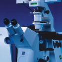
Confocal Microscopy – Zeiss LSM 510 Confocal Microscope
Faculty of Science
Core Facility
McGill University
The Zeiss LSM 510 is a high-resolution laser scanning confocal microscope (LSCM) designed for advanced fluorescence imaging and three-dimensional reconstruction of biological and material samples. This system provides precise optical sectioning, enabling in-depth visualization of cellular structures and dynamic processes with exceptional clarity.
Key Features:
✅ Multi-Laser Excitation – Supports multiple fluorescence channels for versatile imaging applications.
✅ High-Resolution Optical Sectioning – Enables detailed 3D reconstruction of cellular and subcellular structures.
✅ Live-Cell Imaging Capability – Compatible with environmental control systems for real-time monitoring of dynamic biological processes.
✅ Advanced Image Processing & Analysis – Integrated software tools for quantification, colocalization analysis, and spectral unmixing.
This system is ideal for cell biology, neuroscience, developmental biology, and materials science applications requiring high-contrast fluorescence imaging.
Facility for Electron Microscopy Research
Faculty of Science
Research lab focused on advancing scientific knowledge and innovation.
JO
Joaquin Ortega
EquipmentAvailable
Confocal Microscopy – Zeiss LSM 510 Confocal Microscope
Faculty of Science
Core Facility
McGill University
The Zeiss LSM 510 is a high-resolution laser scanning confocal microscope (LSCM) designed for advanced fluorescence imaging and three-dimensional reconstruction of biological and material samples. This system provides precise optical sectioning, enabling in-depth visualization of cellular structures and dynamic processes with exceptional clarity.
Key Features:
✅ Multi-Laser Excitation – Supports multiple fluorescence channels for versatile imaging applications.
✅ High-Resolution Optical Sectioning – Enables detailed 3D reconstruction of cellular and subcellular structures.
✅ Live-Cell Imaging Capability – Compatible with environmental control systems for real-time monitoring of dynamic biological processes.
✅ Advanced Image Processing & Analysis – Integrated software tools for quantification, colocalization analysis, and spectral unmixing.
This system is ideal for cell biology, neuroscience, developmental biology, and materials science applications requiring high-contrast fluorescence imaging.
Facility for Electron Microscopy Research
Faculty of Science
Research lab focused on advancing scientific knowledge and innovation.
JO
Joaquin Ortega
You might also like
Discover more resources that could support your research