Digital AssetAvailable
Pediatric Atlases (4.5–18.5y)
Faculty of Medicine and Health Sciences
Biomedical Engineering
McGill University
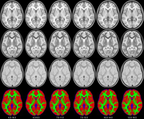
We present an unbiased standard magnetic resonance imaging template brain volume for pediatric data from the 4.5 to 18.5y age range. These volumes were created using data from 324 children enrolled in the NIH-funded MRI study of normal brain development (Almli et al., 2007, Evans and Group 2006). Tools for using these atlases can be found in the Software section.
Methods
The pediatric average atlases are comprised of:
Image pre-processing included non-uniform intensity correction (Sled, 1998) and intensity normalization to a range of 0–100. All T1w MRI data was then transformed into the Talairach-like MNI stereotaxic space using minctracc (Collins, Neelin et al. 1994). Brain masking was performed using BET (Smith, 2002). Age-based subgroups of subjects were created, and all scans within each group were then automatically re-registered to the stereotaxic space using the appropriate template. For each group, an iterative nonlinear co-registration algorithm (Grabner, Janke et al. 2006, Fonov, 2010), was applied to obtain the group averages. The T1-based transformation was then applied to the T2, PD and tissue classified volumes to generate average atlases for these data. Methodological details can be found in (Fonov, 2010).
Viewing
To view the atlases online, click on the appropriate JIV2 link in the Download section below.
Online viewing requires Java browser support. The Java Internet Viewer (JIV2) used here is available for download and personal use under the GNU general public license (GPL).
When viewing, the MNI stereotaxic coordinates (X,Y,Z) are displayed in the first row below the volumes. One can use the left most mouse button to click on any image, and the other cross-sectional images will be updated with the appropriate position. You can also hold the middle/rocker mouse button down while moving up or down, to pan through the image plane. Holding ‘Shift’ with the left or middle button will enable dragging and zooming. When looking at the images, remember that left is left and right is right!
NIST Lab
Faculty of Medicine and Health Sciences
Research lab focused on advancing scientific knowledge and innovation.
LC
Louis Collins
Digital AssetAvailable
Pediatric Atlases (4.5–18.5y)
Faculty of Medicine and Health Sciences
Biomedical Engineering
McGill University
We present an unbiased standard magnetic resonance imaging template brain volume for pediatric data from the 4.5 to 18.5y age range. These volumes were created using data from 324 children enrolled in the NIH-funded MRI study of normal brain development (Almli et al., 2007, Evans and Group 2006). Tools for using these atlases can be found in the Software section.
Methods
The pediatric average atlases are comprised of:
Image pre-processing included non-uniform intensity correction (Sled, 1998) and intensity normalization to a range of 0–100. All T1w MRI data was then transformed into the Talairach-like MNI stereotaxic space using minctracc (Collins, Neelin et al. 1994). Brain masking was performed using BET (Smith, 2002). Age-based subgroups of subjects were created, and all scans within each group were then automatically re-registered to the stereotaxic space using the appropriate template. For each group, an iterative nonlinear co-registration algorithm (Grabner, Janke et al. 2006, Fonov, 2010), was applied to obtain the group averages. The T1-based transformation was then applied to the T2, PD and tissue classified volumes to generate average atlases for these data. Methodological details can be found in (Fonov, 2010).
Viewing
To view the atlases online, click on the appropriate JIV2 link in the Download section below.
Online viewing requires Java browser support. The Java Internet Viewer (JIV2) used here is available for download and personal use under the GNU general public license (GPL).
When viewing, the MNI stereotaxic coordinates (X,Y,Z) are displayed in the first row below the volumes. One can use the left most mouse button to click on any image, and the other cross-sectional images will be updated with the appropriate position. You can also hold the middle/rocker mouse button down while moving up or down, to pan through the image plane. Holding ‘Shift’ with the left or middle button will enable dragging and zooming. When looking at the images, remember that left is left and right is right!
NIST Lab
Faculty of Medicine and Health Sciences
Research lab focused on advancing scientific knowledge and innovation.
LC
Louis Collins
You might also like
Discover more resources that could support your research