EquipmentAvailable
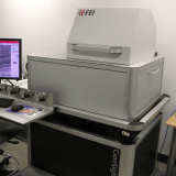
Light Microscopy – FEI CorrSight Fully Automated Correlative Fluorescence Microscope
Faculty of Science
Core Facility
McGill University
The FEI CorrSight is a high-performance, fully automated correlative fluorescence light microscope designed to enhance the efficiency and precision of correlative light and electron microscopy (CLEM) workflows. This system enables seamless integration between fluorescence imaging and electron microscopy, providing researchers with a powerful tool for multi-scale structural and functional analysis.
Key Features:
✅ Correlative Microscopy with MAPS Software – The Modular Automated Processing System (MAPS) facilitates precise correlation between light and electron microscopy data, optimizing image alignment and analysis. Download MAPS Offline Viewer
✅ Dedicated Cryo-Stage – Enables contamination-free imaging of vitrified samples, preserving ultrastructure at high resolution.
✅ Live-Cell Imaging & Automated Fixation – Integrated microfluidics device with an incubated sample area supports real-time imaging, event-triggered chemical fixation, and subsequent processing for electron microscopy.
✅ Fully Automated Workflow – Streamlined automation enhances reproducibility, data consistency, and throughput for CLEM applications.
This system is ideal for researchers in cell biology, structural biology, and materials science requiring high-precision fluorescence imaging for correlative electron microscopy studies.
Facility for Electron Microscopy Research
Faculty of Science
Research lab focused on advancing scientific knowledge and innovation.
JO
Joaquin Ortega
EquipmentAvailable
Light Microscopy – FEI CorrSight Fully Automated Correlative Fluorescence Microscope
Faculty of Science
Core Facility
McGill University
The FEI CorrSight is a high-performance, fully automated correlative fluorescence light microscope designed to enhance the efficiency and precision of correlative light and electron microscopy (CLEM) workflows. This system enables seamless integration between fluorescence imaging and electron microscopy, providing researchers with a powerful tool for multi-scale structural and functional analysis.
Key Features:
✅ Correlative Microscopy with MAPS Software – The Modular Automated Processing System (MAPS) facilitates precise correlation between light and electron microscopy data, optimizing image alignment and analysis. Download MAPS Offline Viewer
✅ Dedicated Cryo-Stage – Enables contamination-free imaging of vitrified samples, preserving ultrastructure at high resolution.
✅ Live-Cell Imaging & Automated Fixation – Integrated microfluidics device with an incubated sample area supports real-time imaging, event-triggered chemical fixation, and subsequent processing for electron microscopy.
✅ Fully Automated Workflow – Streamlined automation enhances reproducibility, data consistency, and throughput for CLEM applications.
This system is ideal for researchers in cell biology, structural biology, and materials science requiring high-precision fluorescence imaging for correlative electron microscopy studies.
Facility for Electron Microscopy Research
Faculty of Science
Research lab focused on advancing scientific knowledge and innovation.
JO
Joaquin Ortega
You might also like
Discover more resources that could support your research