EquipmentAvailable
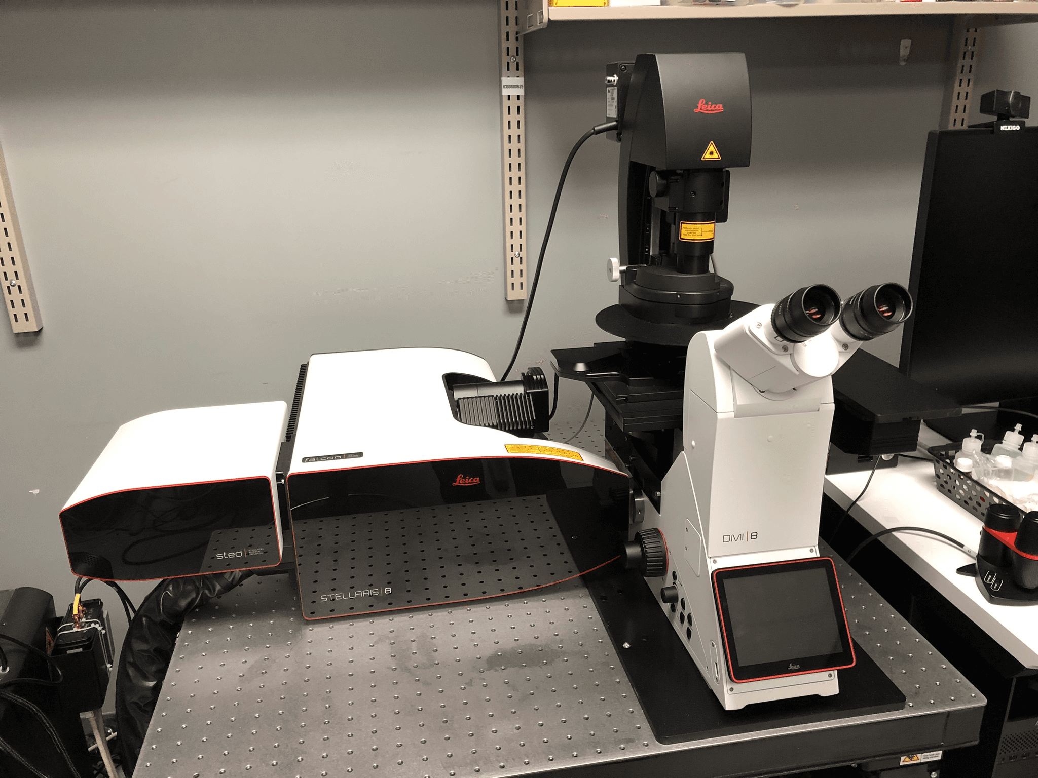
Leica STELLARIS 8 - STED-FLIM
Faculty of Science
Core Facility
McGill University
Stellaris 8 Confocal Microscope
DMi8 motorized and automated inverted microscope stand
STED microscopy and lifetime‐based imaging with tauSense and FALCON (TCSPC‐based FLIM)
Features:
- 4D Imaging, multi-color, z-stack, timelapse
- DIC
- Navigator mode for easy overview and tiling
- LIGHTNING module for enhanced resolution
- STED
- FLIM
- Excitation and emission lambda scans to characterize sample or for spectral unmixing
- Stage‐top incubation system
Light Sources:
- LED3 white light source for epi-fluorescence visualization
- 405nm laser
- Tunable White Light Laser (WLL) with 350 laser lines 440‐790nm (pulsed with 80MHz repetition rate)
- 775nm depletion laser for 3D STED microscopy
Detectors:
- 3 HyD S detectors
- 2 HyD X detectors
Objectives:
PL APO 10x/0.40 CS2
- FWD: 2.56mm
- Coverglass: 0.17mm
- Immersion: dry
PL APO 20x/0.75 CS2
- FWD: 0.62mm
- Coverglass: 0.17mm
- Immersion: dry
PL APO 93x/1.30 GLY motCORR STED WHITE
- FWD: 0.3mm
- Coverglass: 0.14‐0.17mm
- Motorized correction collar (motCORR)
- Immersion: Glycerol
PL APO 100x/1.40 oil STED WHITE
- FWD: 0.13mm
- Coverglass: 0.17mm
- Immersion: oil
Fluorescence Cubes:
15525338 Filter cube LED 405
- Excitation: 405/60
- Emission: 470/40
15525314 Filter Cube GFP
- Excitation: BP 470/40
- Emission: BP 525/50
15525303 Filter Cube RHOD LP
- Excitation: BP 540/45
- Emission: LP 590
Location:
- Located in Bellini Building, Room 151.
Initial training sessions usually last about 3 hours and include the hourly microscope fee plus the technical support fee.
IMPORTANT: ALL training and technical support is by appointment ONLY and will depend on the availability of the ABIF staff.
Advanced BioImaging Facility
Faculty of Science
Research lab focused on advancing scientific knowledge and innovation.
CB
Claire Brown
EquipmentAvailable
Leica STELLARIS 8 - STED-FLIM
Faculty of Science
Core Facility
McGill University
Stellaris 8 Confocal Microscope
DMi8 motorized and automated inverted microscope stand
STED microscopy and lifetime‐based imaging with tauSense and FALCON (TCSPC‐based FLIM)
Features:
- 4D Imaging, multi-color, z-stack, timelapse
- DIC
- Navigator mode for easy overview and tiling
- LIGHTNING module for enhanced resolution
- STED
- FLIM
- Excitation and emission lambda scans to characterize sample or for spectral unmixing
- Stage‐top incubation system
Light Sources:
- LED3 white light source for epi-fluorescence visualization
- 405nm laser
- Tunable White Light Laser (WLL) with 350 laser lines 440‐790nm (pulsed with 80MHz repetition rate)
- 775nm depletion laser for 3D STED microscopy
Detectors:
- 3 HyD S detectors
- 2 HyD X detectors
Objectives:
PL APO 10x/0.40 CS2
- FWD: 2.56mm
- Coverglass: 0.17mm
- Immersion: dry
PL APO 20x/0.75 CS2
- FWD: 0.62mm
- Coverglass: 0.17mm
- Immersion: dry
PL APO 93x/1.30 GLY motCORR STED WHITE
- FWD: 0.3mm
- Coverglass: 0.14‐0.17mm
- Motorized correction collar (motCORR)
- Immersion: Glycerol
PL APO 100x/1.40 oil STED WHITE
- FWD: 0.13mm
- Coverglass: 0.17mm
- Immersion: oil
Fluorescence Cubes:
15525338 Filter cube LED 405
- Excitation: 405/60
- Emission: 470/40
15525314 Filter Cube GFP
- Excitation: BP 470/40
- Emission: BP 525/50
15525303 Filter Cube RHOD LP
- Excitation: BP 540/45
- Emission: LP 590
Location:
- Located in Bellini Building, Room 151.
Initial training sessions usually last about 3 hours and include the hourly microscope fee plus the technical support fee.
IMPORTANT: ALL training and technical support is by appointment ONLY and will depend on the availability of the ABIF staff.
Advanced BioImaging Facility
Faculty of Science
Research lab focused on advancing scientific knowledge and innovation.
CB
Claire Brown
You might also like
Discover more resources that could support your research