EquipmentAvailable
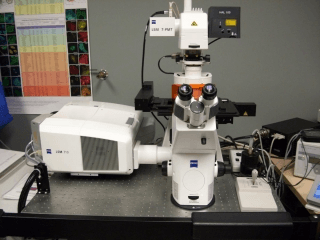
LSM 710 Confocal Laser Scanning Microscope
Faculty of Science
Core Facility
McGill University
LSM 710 Confocal Microscope
Zeiss AxioObserver fully motorized inverted microscope
Features:
- 4D Imaging, multi-color, z-stack timelapse.
- DIC.
- FRET.
- FRAP.
- Meta Spectral imaging.
- Imaging 3D cultures.
- Live cell imaging.
- Tiling.
- Multipoint for timelapse of multiwall plates.
Light Sources:
- X-Cite 120 LED
- 405 nm BLUE DIODE LASER 30 mW
- Ar Ion laser 458/488/514 nm 25 mW
- DPSS-laser 561nm 20mW
- HeNe Red 633 nm 5 mW
Objectives:
- 10X EC PLAN NEOFLUAR, NA=0.30
- 20X PLAN APOCHROMAT, NA=0.80
- 63X PLAN APOCHROMAT, NA=1.4, OIL, DIC
- 100X PLAN APOCHROMAT, NA=1.4, OIL, DIC
Fluorescence Cubes:
- Zeiss FS 49 (DAPI)
- Zeiss FS 38 (eGFP)
- Zeiss FS 45 (mCherry)
- DIC polarizer
Optional
- Texas Red
- YFP
- CFP
Location:
- Located in Bellini Building, Room 151.
Initial training sessions usually last about 3 hours and include the hourly microscope fee plus the technical support fee.
IMPORTANT: ALL training and technical support is by appointment ONLY and will depend on the availability of the ABIF staff.
Advanced BioImaging Facility
Faculty of Science
Research lab focused on advancing scientific knowledge and innovation.
CB
Claire Brown
EquipmentAvailable
LSM 710 Confocal Laser Scanning Microscope
Faculty of Science
Core Facility
McGill University
LSM 710 Confocal Microscope
Zeiss AxioObserver fully motorized inverted microscope
Features:
- 4D Imaging, multi-color, z-stack timelapse.
- DIC.
- FRET.
- FRAP.
- Meta Spectral imaging.
- Imaging 3D cultures.
- Live cell imaging.
- Tiling.
- Multipoint for timelapse of multiwall plates.
Light Sources:
- X-Cite 120 LED
- 405 nm BLUE DIODE LASER 30 mW
- Ar Ion laser 458/488/514 nm 25 mW
- DPSS-laser 561nm 20mW
- HeNe Red 633 nm 5 mW
Objectives:
- 10X EC PLAN NEOFLUAR, NA=0.30
- 20X PLAN APOCHROMAT, NA=0.80
- 63X PLAN APOCHROMAT, NA=1.4, OIL, DIC
- 100X PLAN APOCHROMAT, NA=1.4, OIL, DIC
Fluorescence Cubes:
- Zeiss FS 49 (DAPI)
- Zeiss FS 38 (eGFP)
- Zeiss FS 45 (mCherry)
- DIC polarizer
Optional
- Texas Red
- YFP
- CFP
Location:
- Located in Bellini Building, Room 151.
Initial training sessions usually last about 3 hours and include the hourly microscope fee plus the technical support fee.
IMPORTANT: ALL training and technical support is by appointment ONLY and will depend on the availability of the ABIF staff.
Advanced BioImaging Facility
Faculty of Science
Research lab focused on advancing scientific knowledge and innovation.
CB
Claire Brown
You might also like
Discover more resources that could support your research