EquipmentAvailable
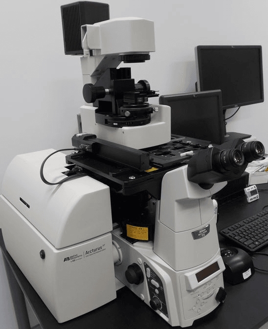
Arcturus XT laser capture microdissection system
Faculty of Medicine and Health Sciences
Research Institute of the McGill University Health Centre
McGill University
The Arcturus XT provides ultimate flexibility in sample source and preparation
The unique system combines Infrared (IR) laser capture and Ultraviolet (UV) laser cutting which can be used on any slide type and sample preparation. Choose from glass, glass membrane or framed membrane slides for contact or non-contact microdissection.
Any specimen preparation may be used:
- Thin or thick sections
- Frozen or formalin-fixed tissues
- Stained, fluorescently stained, or unstained sections
- Hydrated or dehydrated specimens
- Cell cultures
Specifications
Microscope: Nikon Eclipse® Ti-E microscope base. Standard microscope operation outside of LCM operating software
Lasers: IR capture laser: solid-state (810 nm)
Enhanced UV cutting laser: solid-state, diode-pumped Q-switched (345 nm) with adjustable laser current (0–100%) and pulse frequency (10–5,000 Hz)
Illumination: High-intensity LED illumination system
Objectives: 2x, 10x, 20x and 40x Nikon CFI60
Intermediate magnification: 1.5x optical magnification
Stage: Insert for three 75 mm x 25 mm slides, insert for large-format slides; Two positions, adjustable to 75 mm x 25 mm, 75 mm x 38 mm, 75 mm x 50 mm and stage insert for petri dish: 50 mm x 7 mm
Contrast methods: Bright-field , phase contrast (PhL, Ph1, Ph2), differential interference contrast (DIC)
Microdissection camera: 1024 x 768 1/3" color CCD with electronic shutter to 1/10,000s and on-chip integration to 30 seconds
Histopathology Platform - RI-MUHC
Faculty of Medicine and Health Sciences
Research lab focused on advancing scientific knowledge and innovation.
FC
Fazila Chouiali
EquipmentAvailable
Arcturus XT laser capture microdissection system
Faculty of Medicine and Health Sciences
Research Institute of the McGill University Health Centre
McGill University
The Arcturus XT provides ultimate flexibility in sample source and preparation
The unique system combines Infrared (IR) laser capture and Ultraviolet (UV) laser cutting which can be used on any slide type and sample preparation. Choose from glass, glass membrane or framed membrane slides for contact or non-contact microdissection.
Any specimen preparation may be used:
- Thin or thick sections
- Frozen or formalin-fixed tissues
- Stained, fluorescently stained, or unstained sections
- Hydrated or dehydrated specimens
- Cell cultures
Specifications
Microscope: Nikon Eclipse® Ti-E microscope base. Standard microscope operation outside of LCM operating software
Lasers: IR capture laser: solid-state (810 nm)
Enhanced UV cutting laser: solid-state, diode-pumped Q-switched (345 nm) with adjustable laser current (0–100%) and pulse frequency (10–5,000 Hz)
Illumination: High-intensity LED illumination system
Objectives: 2x, 10x, 20x and 40x Nikon CFI60
Intermediate magnification: 1.5x optical magnification
Stage: Insert for three 75 mm x 25 mm slides, insert for large-format slides; Two positions, adjustable to 75 mm x 25 mm, 75 mm x 38 mm, 75 mm x 50 mm and stage insert for petri dish: 50 mm x 7 mm
Contrast methods: Bright-field , phase contrast (PhL, Ph1, Ph2), differential interference contrast (DIC)
Microdissection camera: 1024 x 768 1/3" color CCD with electronic shutter to 1/10,000s and on-chip integration to 30 seconds
Histopathology Platform - RI-MUHC
Faculty of Medicine and Health Sciences
Research lab focused on advancing scientific knowledge and innovation.
FC
Fazila Chouiali
You might also like
Discover more resources that could support your research