EquipmentAvailable
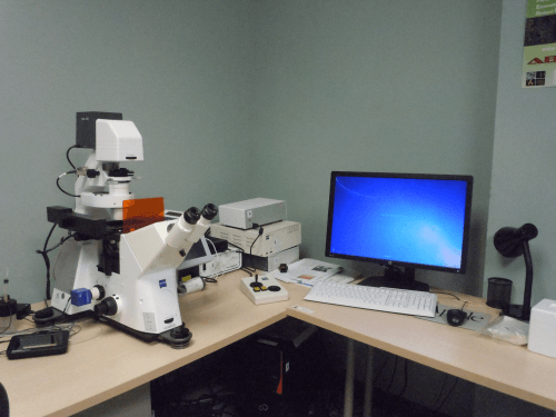
Axiovert 3 - Widefield
Faculty of Science
Core Facility
McGill University
- Zeiss Axio Observer Fully Automated Inverted Microscope.
- Multi-channel epifluorescence, phase contrast, DIC and bright field imaging.
- Z-stack or timelapse.
- Multi-position and tiling.
- Live cell imaging.
Features:
- Zeiss Axiocam 506 monochrome CCD camera 2752 x 2208 pixels, 4.54 um pixels.
- Multi-colour timelapse and z-stacks.
- DIC and Phase contrast.
Light Sources:
- Sola SM solid state light engine.
Objectives:
- 5X N-ACHROPLAN, NA=0.13
- 10X EC PLAN-NEOFLUAR, NA=0.30, Ph1
- 20X LD A-PLAN , NA=0.35, Ph2
- 40X LD A-PLAN, NA=0.55, Ph2
- 63X PLAN-APOCHROMAT, NA=1.4, OIL, DIC
- 100X PLAN-APOCHROMAT, NA=1.4, OIL, Iris
Fluorescence Cubes:
- FS 02 (DAPI)
- FS 24 (FITC)
- FS 15 (TRITC)
- ETmCherry/TR
- FS 32 (Cy5)
Optional
- Texas Red
- YFP
- CFP
Location:
- Located in Bellini Building, Room 141.
Initial training sessions usually last about 3 hours and include the hourly microscope fee plus the technical support fee.
IMPORTANT: ALL training and technical support is by appointment ONLY and will depend on the availability of the ABIF staff.
Advanced BioImaging Facility
Faculty of Science
Research lab focused on advancing scientific knowledge and innovation.
CB
Claire Brown
EquipmentAvailable
Axiovert 3 - Widefield
Faculty of Science
Core Facility
McGill University
- Zeiss Axio Observer Fully Automated Inverted Microscope.
- Multi-channel epifluorescence, phase contrast, DIC and bright field imaging.
- Z-stack or timelapse.
- Multi-position and tiling.
- Live cell imaging.
Features:
- Zeiss Axiocam 506 monochrome CCD camera 2752 x 2208 pixels, 4.54 um pixels.
- Multi-colour timelapse and z-stacks.
- DIC and Phase contrast.
Light Sources:
- Sola SM solid state light engine.
Objectives:
- 5X N-ACHROPLAN, NA=0.13
- 10X EC PLAN-NEOFLUAR, NA=0.30, Ph1
- 20X LD A-PLAN , NA=0.35, Ph2
- 40X LD A-PLAN, NA=0.55, Ph2
- 63X PLAN-APOCHROMAT, NA=1.4, OIL, DIC
- 100X PLAN-APOCHROMAT, NA=1.4, OIL, Iris
Fluorescence Cubes:
- FS 02 (DAPI)
- FS 24 (FITC)
- FS 15 (TRITC)
- ETmCherry/TR
- FS 32 (Cy5)
Optional
- Texas Red
- YFP
- CFP
Location:
- Located in Bellini Building, Room 141.
Initial training sessions usually last about 3 hours and include the hourly microscope fee plus the technical support fee.
IMPORTANT: ALL training and technical support is by appointment ONLY and will depend on the availability of the ABIF staff.
Advanced BioImaging Facility
Faculty of Science
Research lab focused on advancing scientific knowledge and innovation.
CB
Claire Brown
You might also like
Discover more resources that could support your research