Digital AssetAvailable
SEM Imaging of Bacteria or Cells after CPD
Faculty of Science
Core Facility
McGill University
Prepared by Jeannie Mui and S Kelly Sears
Ultracentrifugation protocol
- Collect your samples by ultracentrifugation or other means. For ultracentrifugation, spin as needed, pour off supernatant and add conditioned media until the entire volume is processed into a pellet.
- Wash the pellet for 30 min at 4°C in 2 to 3 mL of ice-cold 0.1 μm sterile-filtered 1X PBS on a shaker, followed by another spin and one final wash/spin to remove secreted proteins and other components.
- Resuspend pellet in 2.5% glutaraldehyde fixative solution in 0.1M sodium cacodylate buffer (final pellet resuspension depends on how much is the starting material, the number of TEM grids you want to use, and the amount of bacteria in your sample, 5 to 10 µL of sample for each TEM grid). The concentration should be around 1 to 5 μg/μL.
- Transfer the bacteria suspension to a 1.5 mL Eppendorf test tube and store them at 4oC.
- Bring to the FEMR for dehydration and critical point drying.
Preparing poly-L-lysine coated coverslips
- A 0.01% (w/v) solution is recommended to coat coverslips with poly-L-lysine. When coating the surface with poly-L-lysine, ensure the entire surface is fully covered.
- After a five-minute incubation, remove the excess solution from the coverslip with filter paper.
- Rinse the coverslip three times with a large volume of distilled water, making sure to rinse thoroughly. The excess poly-L-lysine solution can be toxic to the cell/bacteria.
- Allow the coverslip to dry uncovered at room temperature or in an oven at a gentle heat. If protected from dust, coated coverslips are stable for two weeks at 4 degrees C or one year at -20 degrees C. Tightly wrap the coverslips with Parafilm for storage.
Sample preparation guidelines for SEM: Bacteria/cells in suspension
- Fix the bacteria/cells with 2.5% glutaraldehyde in 0.1M cacodylate buffer for a minimum of 30 to 60 minutes. Fixing at 4oC may improve the preservation of some specimens. To fix, remove most of the media liquid but not all to avoid drying the sample, which adversely affects ultrastructure. Briefly wash with PBS.
- Samples may be fixed again in 1% osmium tetroxide for another 30 to 60 minutes. Post-fixation with osmium tetroxide can be eliminated with an extended primary fixation, i.e., 24-72 hours at 4 degrees C with the primary fixative. Advantage: Less cracking, (caused by shrinkage), during drying process as the samples are less brittle.
- Pipette the bacteria suspension onto a poly-L-lysine coated 12 mm circular glass coverslip (see above) for 15 to 30 minutes. Do not allow the sample to dry.
- Rinse three times using the same buffer for five minutes each rinse (can hold the sample up to 72 hours at 4 degrees C at this point before dehydration)
- Dehydrate the bacteria coverslip with a graded series of ethyl alcohol (ethanol):dH2O, i.e., 30, 50, 70, 80, 90, and 100% (x3) at room temperature; 10 minutes for each is sufficient. Specimens can be stored overnight at 4oC at 70% if necessary, and the dehydration process can be completed the next day. To avoid shrinkage, do not keep the samples in 100% ethanol for an extended period.
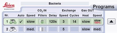
- After the second dehydration with 100% ethanol, immediately transfer the coverslips to the Leica Microsystems EM CPD300 or chemically dry using hexamethyldisilazane (HMDS). Use the recipe below for the CPD dehydration.
Image by Leica Microsystems.
- Sputter coat the coverslips with ~5 nm thickness of platinum using the Leica EM ACE600 sputter coater and image with the Quanta 450 FE-SEM.
Chemically Drying with Hexamethyldisilazane (HMDS)
- Dehydrate the bacteria coverslip with a graded series of ethyl alcohol (ethanol):hexamethyldisilazane (HMDS), i.e., 30, 50, 70, 80, 90, and 100% (x3) at room temperature; two to three hours each step.
Facility for Electron Microscopy Research
Faculty of Science
Research lab focused on advancing scientific knowledge and innovation.
JO
Joaquin Ortega
Digital AssetAvailable
SEM Imaging of Bacteria or Cells after CPD
Faculty of Science
Core Facility
McGill University
Prepared by Jeannie Mui and S Kelly Sears
Ultracentrifugation protocol
- Collect your samples by ultracentrifugation or other means. For ultracentrifugation, spin as needed, pour off supernatant and add conditioned media until the entire volume is processed into a pellet.
- Wash the pellet for 30 min at 4°C in 2 to 3 mL of ice-cold 0.1 μm sterile-filtered 1X PBS on a shaker, followed by another spin and one final wash/spin to remove secreted proteins and other components.
- Resuspend pellet in 2.5% glutaraldehyde fixative solution in 0.1M sodium cacodylate buffer (final pellet resuspension depends on how much is the starting material, the number of TEM grids you want to use, and the amount of bacteria in your sample, 5 to 10 µL of sample for each TEM grid). The concentration should be around 1 to 5 μg/μL.
- Transfer the bacteria suspension to a 1.5 mL Eppendorf test tube and store them at 4oC.
- Bring to the FEMR for dehydration and critical point drying.
Preparing poly-L-lysine coated coverslips
- A 0.01% (w/v) solution is recommended to coat coverslips with poly-L-lysine. When coating the surface with poly-L-lysine, ensure the entire surface is fully covered.
- After a five-minute incubation, remove the excess solution from the coverslip with filter paper.
- Rinse the coverslip three times with a large volume of distilled water, making sure to rinse thoroughly. The excess poly-L-lysine solution can be toxic to the cell/bacteria.
- Allow the coverslip to dry uncovered at room temperature or in an oven at a gentle heat. If protected from dust, coated coverslips are stable for two weeks at 4 degrees C or one year at -20 degrees C. Tightly wrap the coverslips with Parafilm for storage.
Sample preparation guidelines for SEM: Bacteria/cells in suspension
- Fix the bacteria/cells with 2.5% glutaraldehyde in 0.1M cacodylate buffer for a minimum of 30 to 60 minutes. Fixing at 4oC may improve the preservation of some specimens. To fix, remove most of the media liquid but not all to avoid drying the sample, which adversely affects ultrastructure. Briefly wash with PBS.
- Samples may be fixed again in 1% osmium tetroxide for another 30 to 60 minutes. Post-fixation with osmium tetroxide can be eliminated with an extended primary fixation, i.e., 24-72 hours at 4 degrees C with the primary fixative. Advantage: Less cracking, (caused by shrinkage), during drying process as the samples are less brittle.
- Pipette the bacteria suspension onto a poly-L-lysine coated 12 mm circular glass coverslip (see above) for 15 to 30 minutes. Do not allow the sample to dry.
- Rinse three times using the same buffer for five minutes each rinse (can hold the sample up to 72 hours at 4 degrees C at this point before dehydration)
- Dehydrate the bacteria coverslip with a graded series of ethyl alcohol (ethanol):dH2O, i.e., 30, 50, 70, 80, 90, and 100% (x3) at room temperature; 10 minutes for each is sufficient. Specimens can be stored overnight at 4oC at 70% if necessary, and the dehydration process can be completed the next day. To avoid shrinkage, do not keep the samples in 100% ethanol for an extended period.
- After the second dehydration with 100% ethanol, immediately transfer the coverslips to the Leica Microsystems EM CPD300 or chemically dry using hexamethyldisilazane (HMDS). Use the recipe below for the CPD dehydration.
Image by Leica Microsystems.
- Sputter coat the coverslips with ~5 nm thickness of platinum using the Leica EM ACE600 sputter coater and image with the Quanta 450 FE-SEM.
Chemically Drying with Hexamethyldisilazane (HMDS)
- Dehydrate the bacteria coverslip with a graded series of ethyl alcohol (ethanol):hexamethyldisilazane (HMDS), i.e., 30, 50, 70, 80, 90, and 100% (x3) at room temperature; two to three hours each step.
Facility for Electron Microscopy Research
Faculty of Science
Research lab focused on advancing scientific knowledge and innovation.
JO
Joaquin Ortega
You might also like
Discover more resources that could support your research