EquipmentAvailable
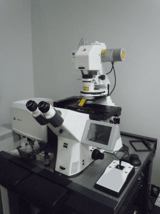
LSM 800 - Confocal Laser Scanning
Faculty of Science
Core Facility
McGill University
- LSM 800 Confocal Microscope
- Zeiss AxioObserver Z.1 fully motorized inverted microscope
Features:
- Multi-color z-stack images
- Preview Function for Tiling
- Multi-point for multi-well plates
Light Sources:
- X-Cite 120 LED
- Halogen
- 405 nm diode laser
- 488 nm diode laser
- 561 nm diode laser
- 640 nm diode laser
Detectors:
- 2 channel multialkali (MA) PMTs
- Transmitted PMT (T-PMT)
- Gallium arsenide phosphide (GaAsP) PMT
Objectives:
- 10X PLAN NEOFLUAR, NA = 0.30
- 20X PLAN APOCHROMAT, NA = 0.80 Ph2
- 40X PLAN NEOFLUAR, NA = 1.30, OIL
- 63X PLAN APOCHROMAT, NA = 1.40, OIL, DIC
Fluorescence Cubes:
- Zeiss FS 49 (DAPI)
- Zeiss FS 13 (GFP)
- Zeiss FS 15 (TRITC)
Location:
- Located in Goodman Cancer Research Centre (GCRC), Room 512.
Initial training sessions usually last about 3 hours and include the hourly microscope fee plus the technical support fee.
IMPORTANT: ALL training and technical support is by appointment ONLY and will depend on the availability of the ABIF staff.
Advanced BioImaging Facility
Faculty of Science
Research lab focused on advancing scientific knowledge and innovation.
CB
Claire Brown
EquipmentAvailable
LSM 800 - Confocal Laser Scanning
Faculty of Science
Core Facility
McGill University
- LSM 800 Confocal Microscope
- Zeiss AxioObserver Z.1 fully motorized inverted microscope
Features:
- Multi-color z-stack images
- Preview Function for Tiling
- Multi-point for multi-well plates
Light Sources:
- X-Cite 120 LED
- Halogen
- 405 nm diode laser
- 488 nm diode laser
- 561 nm diode laser
- 640 nm diode laser
Detectors:
- 2 channel multialkali (MA) PMTs
- Transmitted PMT (T-PMT)
- Gallium arsenide phosphide (GaAsP) PMT
Objectives:
- 10X PLAN NEOFLUAR, NA = 0.30
- 20X PLAN APOCHROMAT, NA = 0.80 Ph2
- 40X PLAN NEOFLUAR, NA = 1.30, OIL
- 63X PLAN APOCHROMAT, NA = 1.40, OIL, DIC
Fluorescence Cubes:
- Zeiss FS 49 (DAPI)
- Zeiss FS 13 (GFP)
- Zeiss FS 15 (TRITC)
Location:
- Located in Goodman Cancer Research Centre (GCRC), Room 512.
Initial training sessions usually last about 3 hours and include the hourly microscope fee plus the technical support fee.
IMPORTANT: ALL training and technical support is by appointment ONLY and will depend on the availability of the ABIF staff.
Advanced BioImaging Facility
Faculty of Science
Research lab focused on advancing scientific knowledge and innovation.
CB
Claire Brown
You might also like
Discover more resources that could support your research