EquipmentAvailable
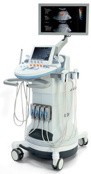
SuperSonic Imagine Aixplorer system
Concordia University
Primary Function: The SuperSonic Imagine Aixplorer system is a cart-based ultrasound imaging system designed for a wide range of diagnostic and research applications, leveraging advanced UltraFast™ imaging technology.
Manufacturer: Originally developed by SuperSonic Imagine (based in Aix-en-Provence, France), the company was acquired by Hologic in 2019. Newer generations are often referred to as Hologic SuperSonic Imagine Aixplorer MACH series.
Technical Specifications:
- Cart-based ultrasound imaging system with an UltraFast™ imaging platform, capable of acquiring images up to 200 times faster than conventional systems.
- Features a scan converter and can be coupled to a variety of linear, curved, micro-convex, and motorized linear array transducers, including new transducer families with pinless connectors.
- Images are displayed on an LCD monitor.
- Adjustable control panel with an integrated touch screen (SonicPad™) for quick and efficient ultrasound exams, designed for improved workflow and user experience (ALARA principles).
- Allows for comprehensive measurements and associated calculations, including cardiac, vascular, OB/GYN, and general calculations.
- Capability to capture images to digital memory or to an external device.
- Functionality to review diagnostic studies in the form of a report.
- Conforms to the DICOM 2018b standard.
- Ergonomic design includes larger monitors, tiltable touch panel, motorized height adjustment, and flexible control panel movement.
Imaging Modes:
- B-Mode (with enhanced penetration, tissue contrast, and spatial resolution)
- M-mode
- Color Doppler imaging (with improved sensitivity and frame rates)
- Pulsed Wave Doppler
- Harmonic Imaging
- Amplitude Power Doppler imaging
- Directional Amplitude Power imaging
- Contrast Imaging (CEUS, optional on specific transducers)
- Elasticity Imaging (ShearWave™ Elastography (SWE™) / ShearWave™ PLUS for real-time tissue stiffness quantification, up to 1,200 kPa or 20 m/s)
- 3D imaging
- Angio PL.U.S. (PLanewave UltraSensitive Imaging for microvascularization)
- TriVu (real-time simultaneous imaging combining B-mode, SWE, and Color+)
- Needle PL.U.S. (for simultaneous visualization of anatomical structures and biopsy needles)
Imaging Applications:
- Abdominal (including advanced liver disease assessment for shear wave speed, tissue stiffness, brightness ratio, vascularization, and perfusion)
- Small Organs
- Musculoskeletal
- Superficial Musculoskeletal
- Vascular
- Peripheral Vascular
- OB-GYN
- Pelvic
- Pediatric
- Neonatal/Adult Cephalic
- Urology
- Intraoperative
- Transrectal
- Transvaginal
- Non-invasive Cardiac
- Breast (including breast lesion diagnosis, characterization, biopsy planning, and therapy monitoring)
- Thyroid
- Prostate
Applications: Comprehensive ultrasound diagnostics, research across multiple anatomical regions, with a strong focus on non-invasive assessment of tissue stiffness for conditions like liver fibrosis and breast lesions.
PERFORM Centre | School of Health
Research lab focused on advancing scientific knowledge and innovation.
WK
Wendy Kunin
EquipmentAvailable
SuperSonic Imagine Aixplorer system
Concordia University
Primary Function: The SuperSonic Imagine Aixplorer system is a cart-based ultrasound imaging system designed for a wide range of diagnostic and research applications, leveraging advanced UltraFast™ imaging technology.
Manufacturer: Originally developed by SuperSonic Imagine (based in Aix-en-Provence, France), the company was acquired by Hologic in 2019. Newer generations are often referred to as Hologic SuperSonic Imagine Aixplorer MACH series.
Technical Specifications:
- Cart-based ultrasound imaging system with an UltraFast™ imaging platform, capable of acquiring images up to 200 times faster than conventional systems.
- Features a scan converter and can be coupled to a variety of linear, curved, micro-convex, and motorized linear array transducers, including new transducer families with pinless connectors.
- Images are displayed on an LCD monitor.
- Adjustable control panel with an integrated touch screen (SonicPad™) for quick and efficient ultrasound exams, designed for improved workflow and user experience (ALARA principles).
- Allows for comprehensive measurements and associated calculations, including cardiac, vascular, OB/GYN, and general calculations.
- Capability to capture images to digital memory or to an external device.
- Functionality to review diagnostic studies in the form of a report.
- Conforms to the DICOM 2018b standard.
- Ergonomic design includes larger monitors, tiltable touch panel, motorized height adjustment, and flexible control panel movement.
Imaging Modes:
- B-Mode (with enhanced penetration, tissue contrast, and spatial resolution)
- M-mode
- Color Doppler imaging (with improved sensitivity and frame rates)
- Pulsed Wave Doppler
- Harmonic Imaging
- Amplitude Power Doppler imaging
- Directional Amplitude Power imaging
- Contrast Imaging (CEUS, optional on specific transducers)
- Elasticity Imaging (ShearWave™ Elastography (SWE™) / ShearWave™ PLUS for real-time tissue stiffness quantification, up to 1,200 kPa or 20 m/s)
- 3D imaging
- Angio PL.U.S. (PLanewave UltraSensitive Imaging for microvascularization)
- TriVu (real-time simultaneous imaging combining B-mode, SWE, and Color+)
- Needle PL.U.S. (for simultaneous visualization of anatomical structures and biopsy needles)
Imaging Applications:
- Abdominal (including advanced liver disease assessment for shear wave speed, tissue stiffness, brightness ratio, vascularization, and perfusion)
- Small Organs
- Musculoskeletal
- Superficial Musculoskeletal
- Vascular
- Peripheral Vascular
- OB-GYN
- Pelvic
- Pediatric
- Neonatal/Adult Cephalic
- Urology
- Intraoperative
- Transrectal
- Transvaginal
- Non-invasive Cardiac
- Breast (including breast lesion diagnosis, characterization, biopsy planning, and therapy monitoring)
- Thyroid
- Prostate
Applications: Comprehensive ultrasound diagnostics, research across multiple anatomical regions, with a strong focus on non-invasive assessment of tissue stiffness for conditions like liver fibrosis and breast lesions.
PERFORM Centre | School of Health
Research lab focused on advancing scientific knowledge and innovation.
WK
Wendy Kunin
You might also like
Discover more resources that could support your research