Gladstone Institutes
Flow Cytometry Core
Sort cells on up to 18 colors or run large spectral panels, with the staff or on your own.
1650 Owens Street, San Francisco, CA 94158
The core
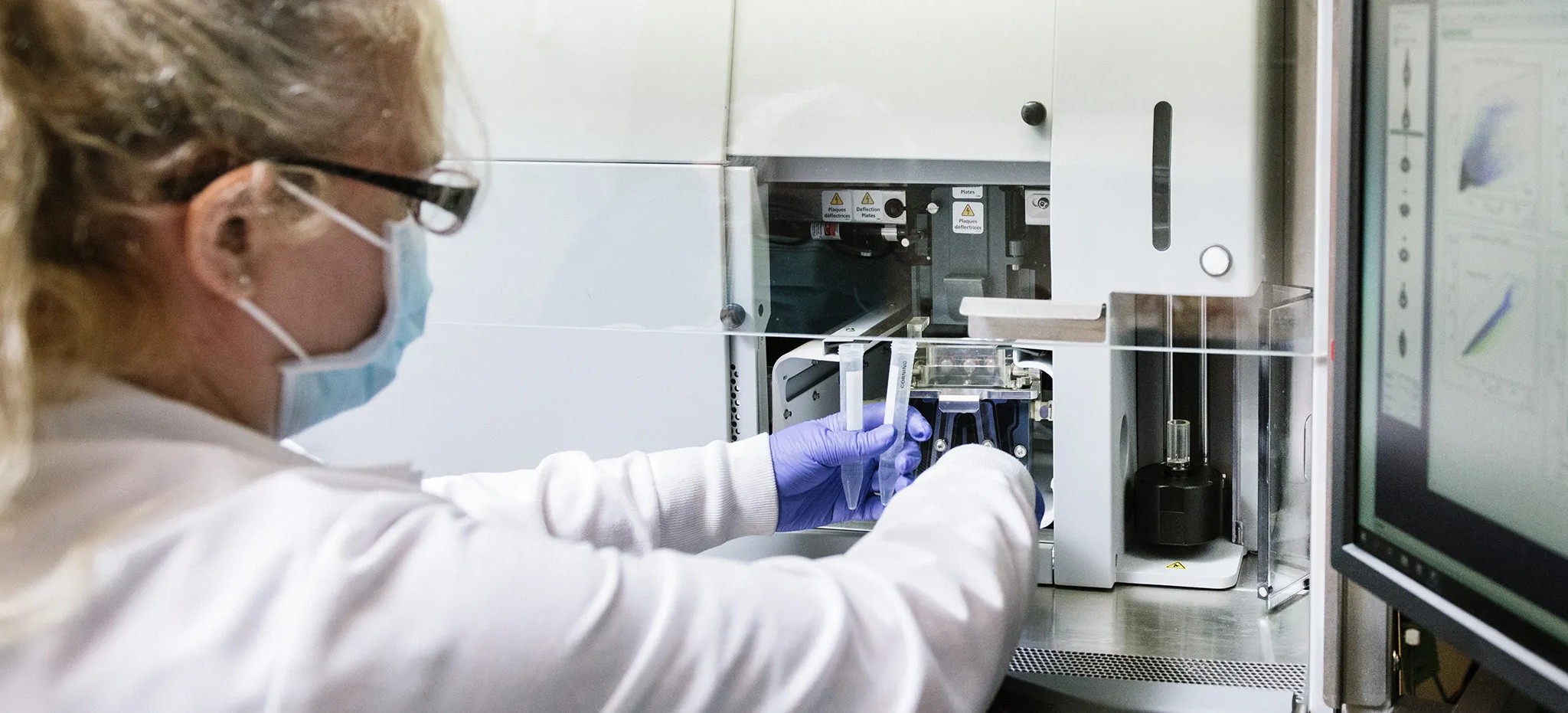
The Flow Cytometry Core analyzes and isolates cells by their phenotype or their function. Sort on up to 18 fluorescent colors, in bulk or one cell per well, or read large panels in a single tube on the spectral analyzer.
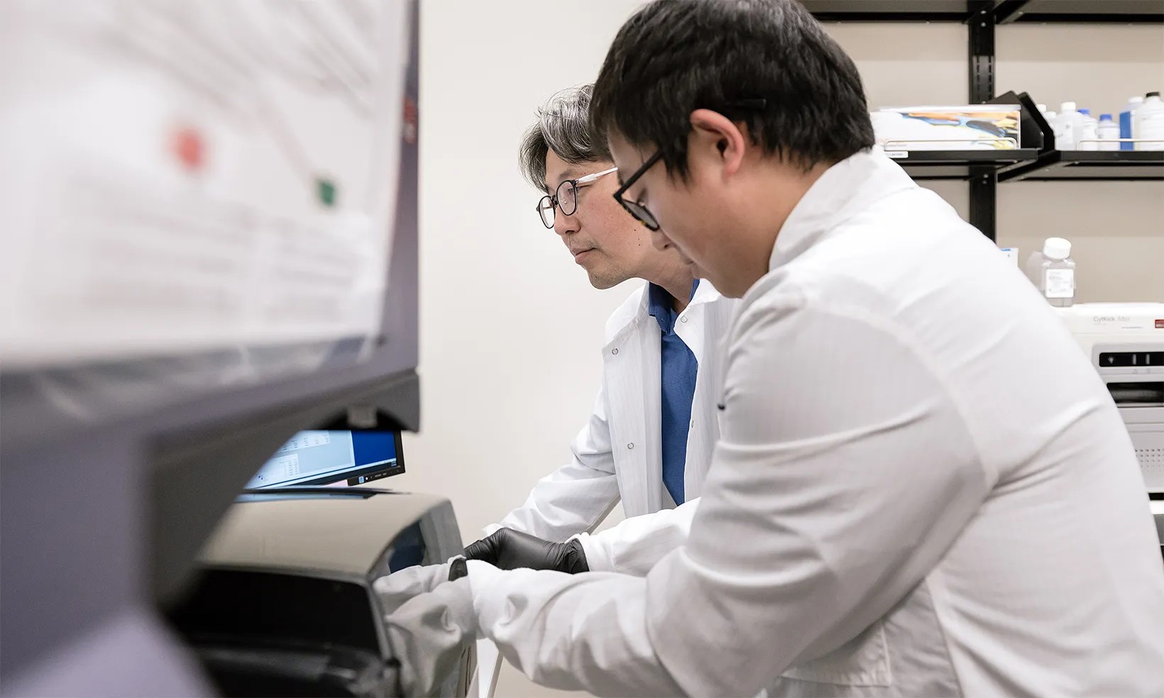
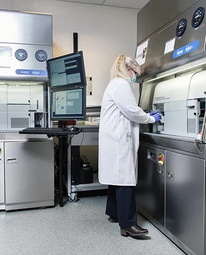
There are two ways to work with the core. Hand your samples to the staff, who can stain them, run them and analyze the data. Or train with them and run the analyzers and sorters yourself, after hours too, once you have shown you can run, maintain and troubleshoot the instrument.
The staff design experiments and antibody panels with you, and the core can work alongside the other Gladstone cores as one pipeline for studying cells.
What you can measure
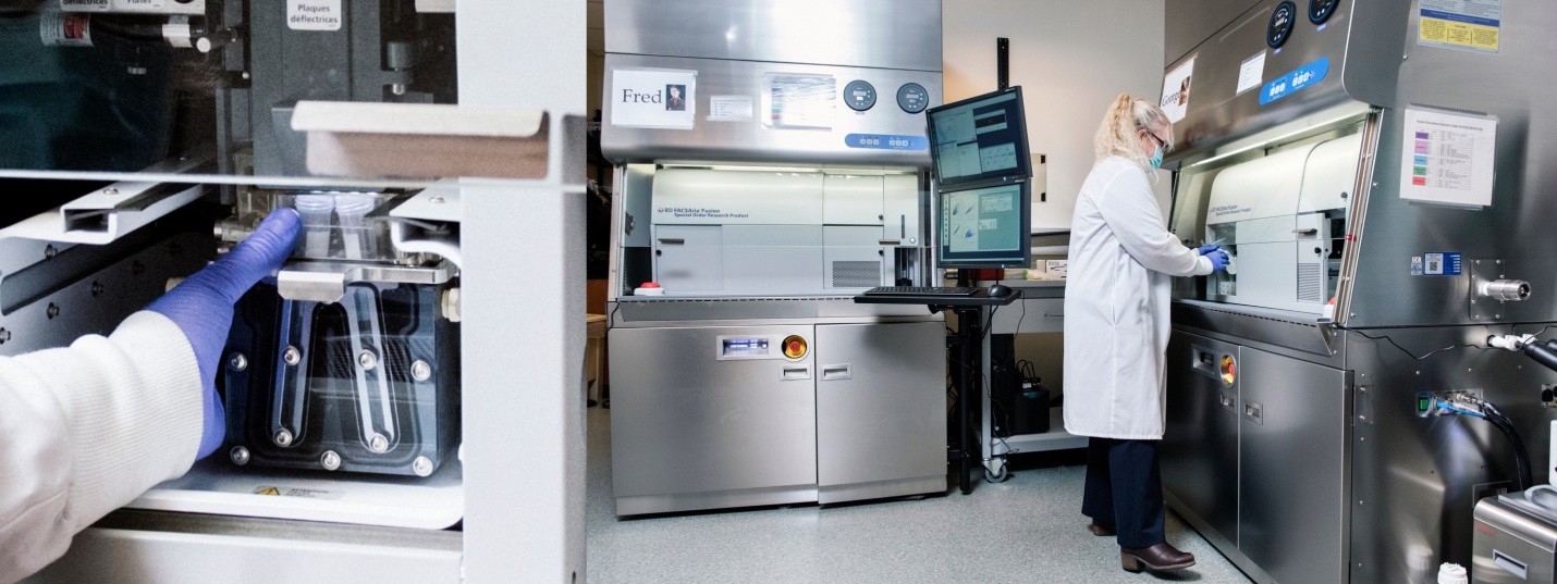
Cell sorting. Up to 18 fluorescent parameters on five lasers: 355, 405, 488, 561 and 640 nm. Bulk, single-cell and index sorting into tubes, any plate from 12 to 384 wells, Terasaki plates or slides. Single cells go into 96-well plates for cloning or sequencing. Aseptic sorting is available.
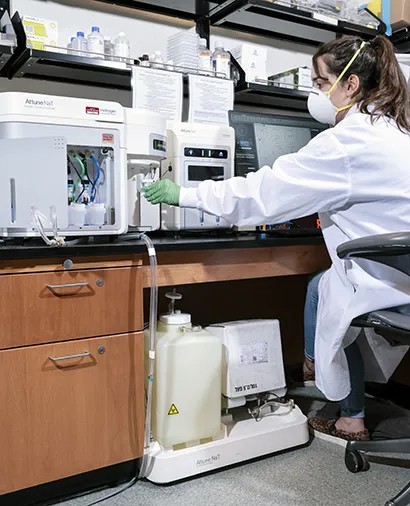
Analysis. Surface, intracellular and functional markers and fluorescent proteins, including phospho-flow, cell cycle and mitochondrial assays. Three analyzers run 96- and 384-well plates.
Imaging cytometry. A picture of every cell at 20x, 40x or 60x, for co-localization, internalization, cell cycle and mitosis, signaling, shape change and nuclear translocation.
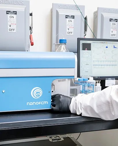
Nanoparticles. Size, concentration and fluorescence of single particles from about 40 nm to 1 micron: extracellular vesicles, mitochondria, bacteria, viruses and synthetic nanoparticles.
iPS cell pluripotency. A marker panel that tells you whether your human iPS cells are pluripotent, with results in about a day.
Before you bring samples
A single-cell suspension. Any cells between 500 nm and 20 µm, in 5 or 15 ml Falcon tubes, Eppendorf tubes, multi-well plates or on slides.
Up to 5 million cells per ml in PBS or media. Keep serum at 2 percent or less: more can affect the charge on the stream. HEPES (25 mM), EDTA (1 mM) or DNase (10 U/ml) keep cells from clumping. Filter through a cell strainer, and bring extra media in case the sample needs diluting.
A copy of your data. The core saves a copy of your FCS files to an internal server.
Science that ran through the core
Papers that used the core span HIV latency, Alzheimer's disease, aging and stem cells. A few examples:
In vivo chimeric Alzheimer's disease modeling of apolipoprotein E4 toxicity in human neurons. Cell Reports, 2020.
SIRT1 is downregulated by autophagy in senescence and ageing. Nature Cell Biology, 2020.
FOXO1 promotes HIV latency by suppressing ER stress in T cells. Nature Microbiology, 2020.
A molecular mechanism for probabilistic bet hedging and its role in viral latency. PNAS, 2020.
The Publications tab lists more, each with the service it used.
Start a project
Pick a service and press Request service. Say what cells you have and what you want to find or isolate. The staff come back to plan the run with you and send a quote.
To run the instruments yourself, press Request training on any of them. You read and sign the core's general regulations, answer a short questionnaire about your experience, then train on the instrument.
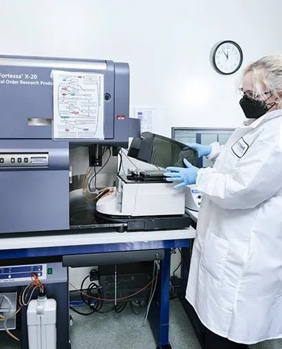
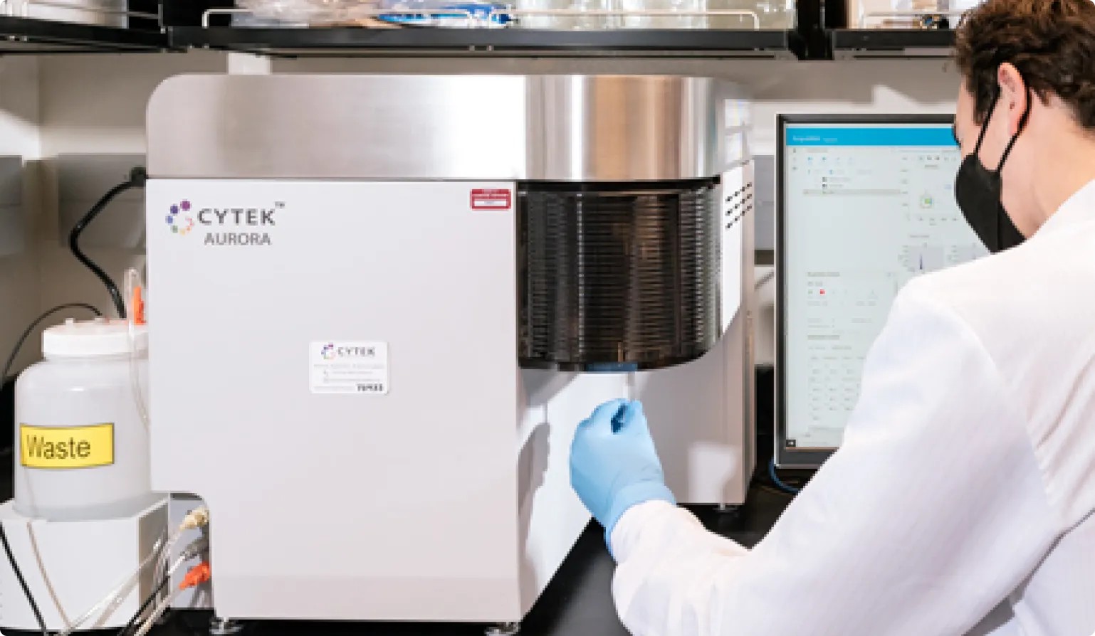
Plates or tubes. The Aurora, LSRFortessa X-20 and Attune NxT all read 96- and 384-well plates.
Contact the facility
Talk to the team about your research, access, or a visit.
Before your first run
New to the core? Write to the staff before you come in. Training starts with the core's general regulations and a short questionnaire about your experience with flow.