Gladstone Institutes
Histology and Light Microscopy Core
From fixed tissue to stained slides and 3D images: histology, confocal, light-sheet and image analysis.
1650 Owens Street, San Francisco, CA 94158
The core
The Histology and Light Microscopy Core helps with every step of an imaging experiment: the design, the sample preparation, the imaging, and the processing and analysis of the images. Bring your tissue for the staff to process, or train with them to use some of the equipment on your own time.
The core serves researchers at Gladstone and at other institutions and companies.
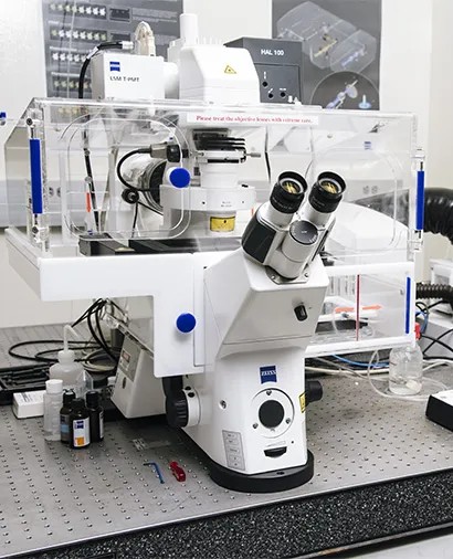
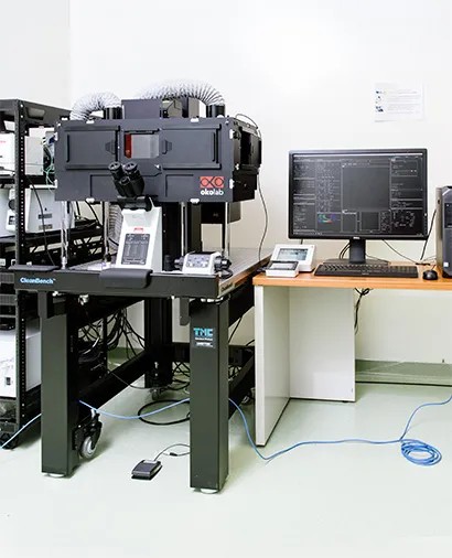
Most of its microscopes go by name. Snow Leopard is an Olympus FV3000 confocal, Tarsier a Zeiss LSM 880 with Airyscan, and the three Coyotes are Keyence widefield microscopes.
What the core does
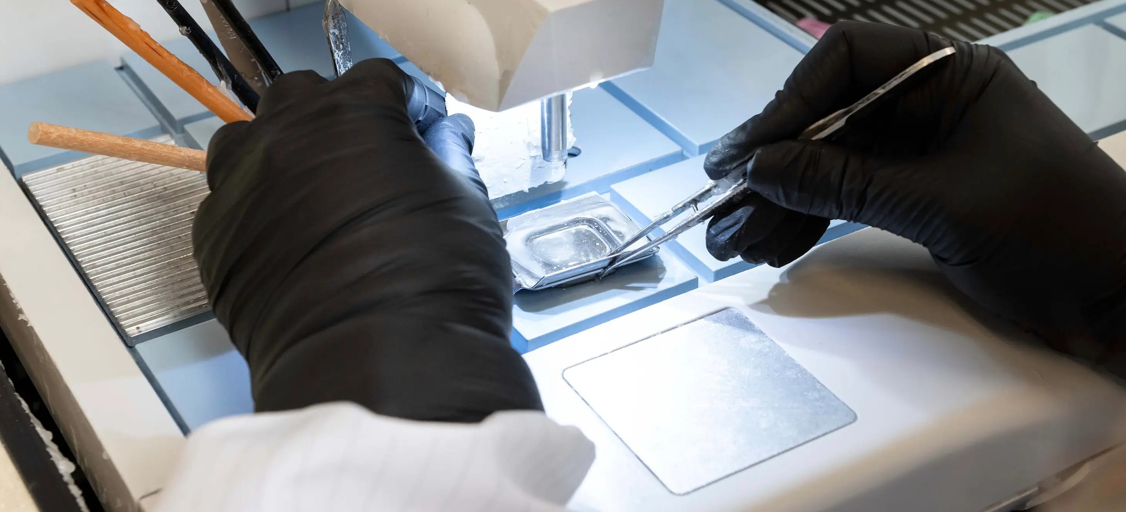
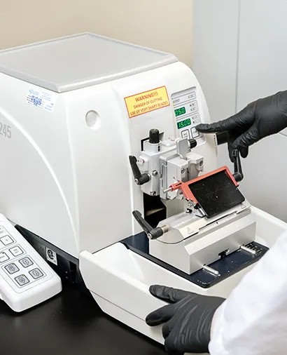
Histology. Processing of paraffin (FFPE) and frozen tissue, embedding, sectioning, staining and whole slide scanning. Stains include H&E, in situ hybridization, TUNEL, and special stains: PAS, Oil Red O, Congo red and Masson's trichrome.
Spatial biology. 10x Visium, run with the Genomics Core.
Microscopy, live and fixed. Confocal and super-resolution, light-sheet with tissue clearing for volumetric imaging, automated whole slide scanning, and widefield.
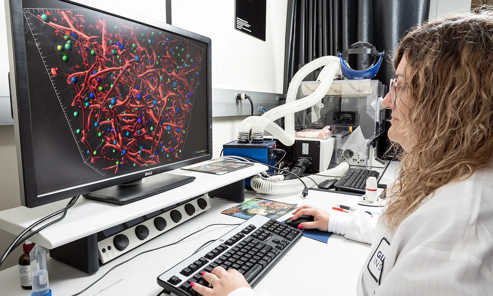
Image analysis. Imaris on the Mammoth workstation in room 327, ImageScope, ImageJ and FIJI, OlyVia, and a workstation of the Gladstone Institute of Cardiovascular Disease.
From a cleared sample to a 3D map
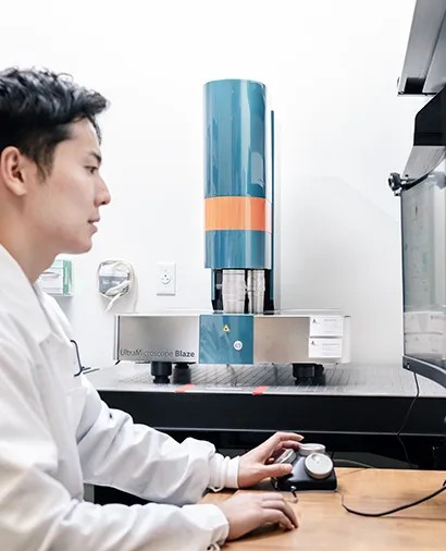
The UltraMicroscope Blaze images cleared samples in 3D, from organoids to a whole adult mouse, quickly and with little photobleaching. Then analyze the volumes in Imaris on the core's Mammoth workstation.
Bringing samples
Fix first. All tissue should be fixed before it comes in. Most tissue does well with fresh perfusion, then 4 percent PFA or 10 percent formalin overnight in the fridge. Brain and bone need longer.
For paraffin, rinse three times in 1x PBS after fixation and store in 70 percent ethanol, ideally stepping up through 25 and 50 percent first.
For frozen sections, bring tissue in 30 percent sucrose on ice, or in OCT blocks or on slides on dry ice. For Visium, ask the core before you prepare anything.
Drop-off and pick-up are in person, in room 367, between 10 am and 3 pm. The core tells you when your samples are ready.
Turnaround is usually 15 to 20 business days from drop-off, once the request is complete. Rush orders take 4 business days. Large or complex projects, or a busy queue, can take longer.
Science that ran through the core
Papers that used the core span neurodegeneration, autism, heart development and human fetal development. A few examples:
Tau reduction prevents key features of autism in mouse models. Neuron, 2020.
Microglial microRNAs mediate sex-specific responses to tau pathology. Nature Neuroscience, 2020.
Single-cell determination of cardiac microtissue structure and function using light sheet microscopy. Tissue Engineering Part C, 2020.
Single-cell analysis of cardiogenesis reveals basis for organ-level developmental defects. Nature, 2019.
The Publications tab lists more, each with the service it used.
Start a project
Talk to the staff before you prepare samples, so your reagents match the core's equipment.
Pick a service and press Request service. Describe the tissue and what you need, and the core comes back with a plan and a quote. Researchers at other nonprofits and at companies sign the core's lab service agreement before the first project. Payment is by purchase order.
To use equipment yourself, press Request training on the instrument. Some of the equipment is open to trained users once the staff grant access. Everyone completes an initial training session before using equipment in the core. Training and staff support are charged by the hour.
Credit the core. The core follows the ABRF authorship guidelines: cite it when your work used its space, equipment, reagents or services, or when its staff helped design or run the experiments.
Contact the facility
Talk to the team about your research, access, or a visit.
Drop-off and pick-up
Bring samples in person to room 367, between 10 am and 3 pm. Technical questions go to the address here; quotes, purchase orders and agreements to [email protected].